DISCOVER NEW FACETS OF VISION
The PENTAX Medical Ultrasound Video Bronchoscope EB19-J10U features crystal clear ultrasound imaging to support diagnostic accuracy, contributes to a smooth facilitation of the EBUS-TBNA, and is ergonomically designed for ease of operation and high working comfort.
An unmatched solution for Endobronchial Ultrasound
Endobronchial Ultrasound (EBUS) and EBUS-guided Transbronchial Needle Aspiration (EBUS-TBNA) has been incorporated into the clinical routine as the gold standard procedure, replacing more invasive methods, for staging lung cancer and evaluating other pulmonary diseases.
The unmatched combination of PENTAX Medical EB19-J10U with ultrasound systems provides a unique solution for accurate diagnosis and staging of lung diseases. State-of-the-art technology ensures the highest imaging performance with crystal clear image quality for precise visualization of the airway wall, surrounding lymph nodes, and adjacent structures.
Leveraging strong synergies
PENTAX Medical provides a combined clinical solution for physicians to clearly visualise and increase the detection of abnormalities, successfully enabling endosonographical diagnostic and therapeutic procedures to the benefit of patients.
Strong synergies between products, highly-specialised application expertise, professional training and education, as well as expert service combine into a unique solution to meet customers’ sophisticated requirements.
Outstanding image quality
Clear ultrasound and endoscopic images result
in improved sensitivity for diagnosis.
Advanced imaging modalities
Advanced imaging modalities contribute
additional diagnostic information.
Portfolio of ultrasound systems
The largest range of EBUS/EUS compatible ultrasound systems – from budget class to high specialisation.
Best clinical
outcomes
Customisation and image optimisation
Image settings can be optimised for each individual user in real-time on-site.
Complementary trans-thoracic probes
The wide range of additional ultrasound probes for complementary imaging in pulmonology departments
completes the EBUS solution.
Training and education
Training programs provide the highest standard of professional education across a range of skill levels, from basic to expert.
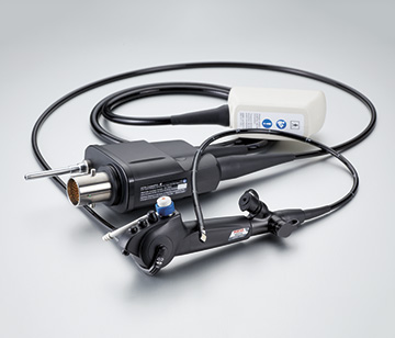
EB19-J10U Ultrasound Video Bronchoscope
The PENTAX Medical Ultrasound Video Bronchoscope EB19-J10U is designed to improve diagnostic outcomes. It features crystal clear ultrasound imaging to support diagnostic accuracy, contributes to a smooth facilitation of the EBUS-TBNA, and is ergonomically designed for ease of operation and high working comfort.
Crystal clear imaging for precise visualisation
Offers state-of-the-art ultrasound image quality for clear visualisation of the airway wall, surrounding lymph nodes, and adjacent structures. It supports orientation and navigation in the airways by the sharpened HD endoscopic view.
Reliable tissue acquisition
The combination of an outstanding ultrasound imaging and an enlarged working channel contribute to an accurate and easier real-time ultrasound-guided transbronchial needle aspiration (EBUS-TBNA) for the diagnosis and staging of lung diseases through the use of a wide range of needles.
Unmatched ergonomic design for ease of operation
Optimal working comfort for users and high patient tolerance provided by the small ultrasound transducer, the slim endoscope outer diameter and the unique ergonomics of the control body.
High precision through optimized design
Crystal clear imaging enhancing diagnosis
- Unmatched ultrasound image quality leading to clear visualisation of the airways, surrounding lymph nodes and adjacent anatomies
- Supports diagnosis and staging of lung cancer and other airway diseases
Optimal visualisation and orientation
- Clear endoscopic view supporting orientation and navigation in the airways
- Enhancing user confidence with permanent ultrasound transducer visibility for better guidance during examination
High precision for EBUS-TBNA
- Optimal needle visualisation aids the targeting of lymph nodes and masses
- Precise navigation of EBUS-TBNA needle thanks to optimal penetration angle
- Effortless, more accurate and reliable sampling through enlarged working channel of 2.2 mm that allows smooth needle delivery
Ease of operation
- Easy handling during the procedure and easy reprocessing due to lightweight control body and redesigned connectors
- Small ultrasound transducer and slim endoscope outer diameter enhancing maneuverability and provide optimal working comfort
Reliable for increased user and patient safety
- Endoscope design and single use accessories meet strong market requirements for reprocessing and product hygiene
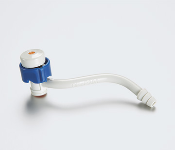
- Single-use suction valve and cleaning brushes to minimize risk of cross-contamination
Enriched diagnostic findings
Broadband Harmonic Imaging
High definition dynamic Tissue Harmonic Imaging (HdTHI) / dynamic Tissue Harmonic Imaging (dTHI)
- Improves spatial resolution and achieves excellent deep area penetration
- Clear boundaries delineation, vascular structures clear of noise and good visualisation of needle tip insertion, are easily achieved with high frame rate
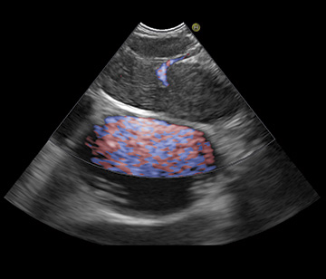
Fine Flow/eFlow
Flow Mapping Technology
- Allows detailed display of blood flow dynamics
- Accurately depicts blood flow dynamics, providing improved spatial resolution and sensitivity at high frame rates
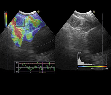
Real-Time Tissue Elastography
(RTE)
- Measures and displays tissue strain in real-time for immediate visualisation of hard and soft tissue
- Helps targeting tissue sampling during EBUS-TBNA procedures
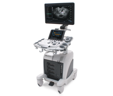
ARIETTA 65PX
Expertly designed to optimise productivity within compact mid-range systems
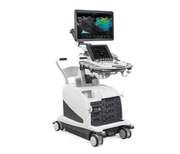
ARIETTA 750PX
High-end ultrasound systems for advanced performance
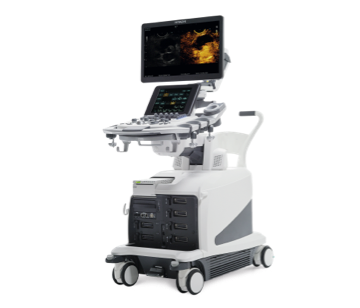
ARIETTA 850PX
The next evolution in ultrasound, designed for high expectations
An innovative pairing: the PENTAX Medical EB19-J10U with DEFINA solution
HD image quality and i-scan technology – at a new level
The compact DEFINA EPK-3000 video processor offers state-of-the-art HD imaging for detection of epithelial changes. This is supported by i-scan, which is a digital image enhancement endoscopy technology and a virtual endoscopy tool. Easily operated with just the touch of a button, it offers rapid imaging results and real-time processing. This i-scan technology guides the user to the area that needs to be biopsied and supports the user in determining the area to be treated.
Superior image quality for better orientation
- Brilliant, high-definition images for a clear, fast, and more detailed visualisation of the mucosal structures
- Dynamic range expansion for excellent far-field illumination and better visibility of the distal area
Supporting diagnostic outcome
- HD image combined with i-scan technology for faster detection, easier demarcation, and support in characterisation
Ease of operation
- Compact design for a better fit in the existing clinical environment
Intended use following recommendation of European guideline
The European guideline recommends the combined use of the EBUS endoscope for endobronchial and esophageal endosonography for the diagnosis and staging of lung cancer.*
In line with this guideline, the intended use of the PENTAX Medical EB19-J10U was expanded to endobronchial and esophageal endosonography which allows the user to perform a combined EBUS-TBNA and EUS-(B)-FNA procedure with only one ultrasound endoscope.
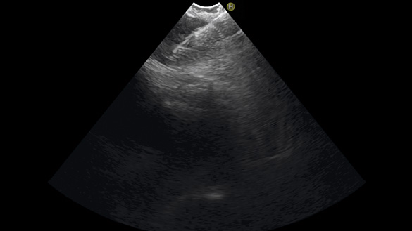
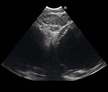
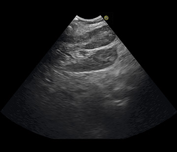
EBUS performed with PENTAX Medical EB19-J10U in the airways showing EBUS-TBNA of lymph node station 7 from right main bronchus.
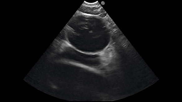
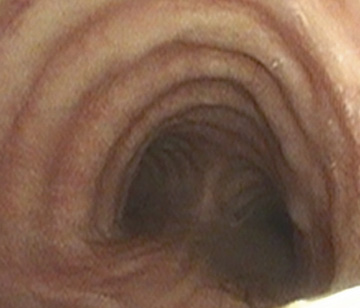
EUS-B esophageal approach using the EBUS endoscope of PENTAX Medical EB19-J10U showing lymph node station 7.
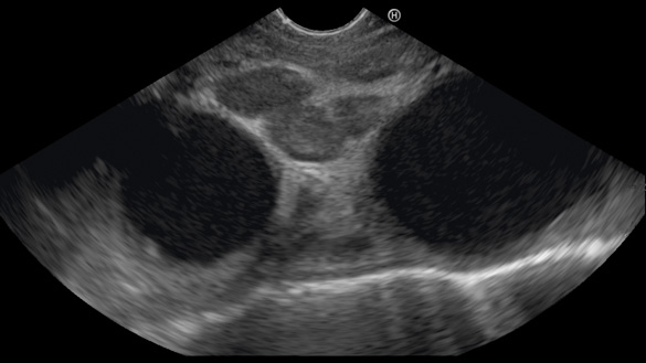
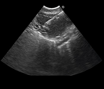
EUS-(B) in the esophagus performed with PENTAX EG-3270UK showing multiple lymph nodes.
For further information about the ESGE, ERS, and ESTS guideline visit:
www.esge.com* “Combined endobronchial and esophageal endosonography for the diagnosis and staging of lung cancer: European Society of Gastrointestinal Endoscopy (ESGE) Guideline, in cooperation with the European Respiratory Society (ERS) and the European Society of Thoracic Surgeons (ESTS)” Peter Vilmann, Paul Frost Clementsen, Sara Colella, Mette Siemsen, Paul De Leyn, Jean-Marc Dumonceau, Felix J. Herth, Alberto Larghi, Enrique Vazquez-Sequeiros, Cesare Hassan, Laurence Crombag, Daniël A. Korevaar, Lars Konge, Jouke T. Annema